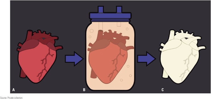
Figure 3. Decellularization schematic. (A) whole heart (may be human, e.g. cadaveric or a transplant reject or, more commonly, from an animal donor of suitable size/ anatomy match, often porcine) is placed into (B) an organ chamber of a decellularization bioreactor and appropriately connected up with suitable tubing and cannulae for perfusion, and the decellularization process initiated. Over a period of time, usually 1 or more days of continuous application of decellularization solution, the heart gradually whitens, indicative of the cell constituent of the tissue being washed away, largely leaving behind the collagen and other connective tissue substance and preserving to a great extent the original organs anatomical architecture with respect to vasculature and parenchyma (C).
Source: Private collection.

