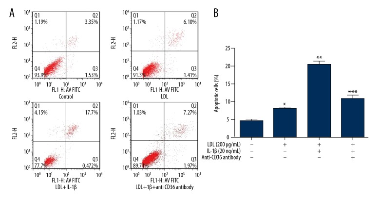
Figure 3.
Treatment with LDL and IL-1β induces apoptosis of MPC5 cells via CD36. MPC5 cells were treated as indicated for 48 h and the percentage of apoptotic cells was determined by flow cytometry. (A) Flow cytometric plots. (B) Percentage of apoptotic cells. * p<0.05 vs. CTR cells; ** p<0.05 vs. CTR cells; *** p<0.05 vs. LDL+IL-1β-treated cells.

