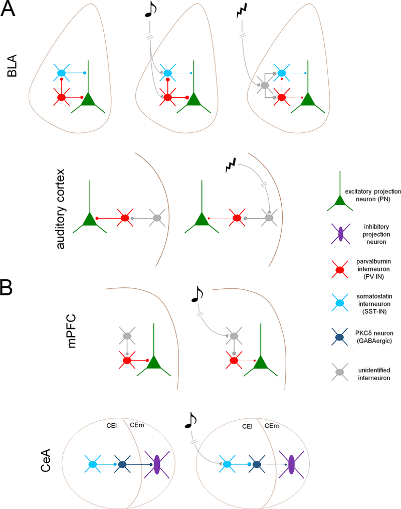
Figure 1. Inhibitory microcircuits for fear memory acquisition (A) and expression (B).

A. Transient disinhibition controls acquisition of aversive conditioning in auditory cortex and basolateral amygdala (BLA). BLA, top. PNs are subject to compartment-specific disinhibition during both CS and US presentation. During CS presentation, PV-INs are excited, inhibiting SST-INs and thereby disinhibiting PN dendrites. During the US, both PV-INs and SST-INs are inhibited by an unidentified cell type. Auditory cortex, bottom. Unconditioned stimulus (US)-mediated acetylcholine release from the basal forebrain excites an undefined interneuron population in cortical layer 1, which suppresses PV-IN firing the thereby disinhibits PNs B. Fear memory expression is mediated by transient disinhibition in the central amygdala (CeA) and medial prefrontal cortex (mPFC). mPFC, top. Following fear conditioning, CS exposure (in the absence of the US) excites an unidentified interneuron population, which suppresses PV-IN firing and thereby disinhibits BLA-projecting excitatory neurons. CeA, bottom. CS presentation excites SST-INs in the lateral division of the central amygdala (CEl), which inhibit PKCδ-expressing GABAergic projection neurons, resulting in disinhibition of brainstem-projecting GABAergic cells in the medial division (CEm).
