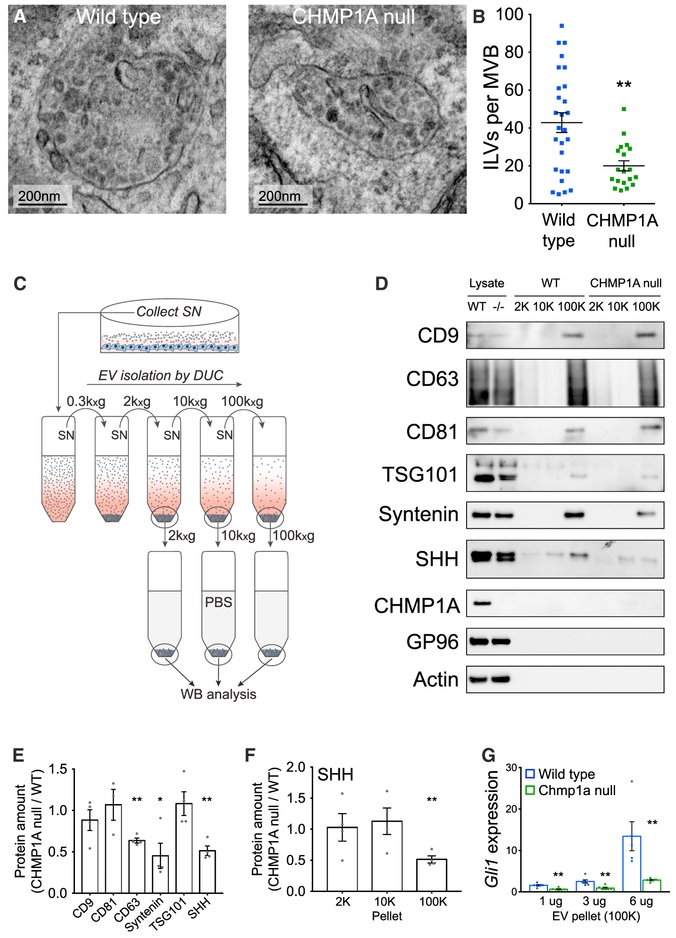
Figure 6. CHMP1A Depletion Decreases Secretion of SHH and Exosomes In Vitro.
(A) Decreased ILV formation in MVBs of CHMP1A null SVG-A cells compared to WT (TEM).
(B) Quantification of (A). Wild-type, n = 27; CHMP1A null, n = 19.
(C) EV isolation procedure by differential ultracentrifugation (DUC) (Kowal et al., 2016).
(D) Representative WB analysis of isolated EVs (2K, 10K, and 100K pellets) from WT and CHMP1A null SVG-A cells expressing SHH. Blot shows EV-specific markers (CD9, CD63, CD81, Syntenin, and TSG101), EV-excluded markers (GP96 and Actin), CHMP1A, and SHH.
(E) Quantification of (D). Wild-type, n = 4; CHMP1A null, n = 4.
(F) Quantification of SHH WB signals in the 2K, 10K, and 100K fractions. Wild-type, n = 4; CHMP1A null, n = 4.
(G) Gli1 induction in NIH 3T3 cells induced by 100K pellet from SHH-transfected SVG-A cells. 1 and 3 μg: wild-type, n = 6; CHMP1A null, n = 6. 6 μg: wild-type, n = 5; CHMP1A null, n = 6.
(B) Mann-Whitney test. (E–G) Two-tailed, unpaired t test. *p < 0.05, **p < 0.01.
Error bars are SEM.

