Figure 6.
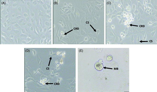
Morphological changes of MDA-MB-231 cells were examined under a phase contrast microscope. Untreated MDA-MB-231 cells (A), MDA-MB-231 cells treated with IC50 concentration of MEDL after 24 h (B), 48 h (C), and 72 h (D) at 200× magnification. MDA-MB-231 cells treated with IC50 concentration of MEDL after 72 h (E) at 400× magnification. (CRD): cell rounding and detachment; (CS): cell shrinkage; (MB): membrane blebbing. Experiment was completed in three independent experiments in triplicate.

