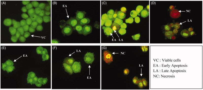
Figure 7.
Fluorescence microscopy examination of MDA-MB-231 cells (400× magnifications). Untreated MDA-MB-231 cells (A). MDA-MB-231 cells treated with IC50 of MEDL after 24 h (B), 48 h (C) and 72 h (D). MDA-MB-231 cells treated with IC50 of 5-FU after 24 h (E), 48 h (F) and 72 h (G). Viable cells displayed the appearance of circular cells, with round and organized nuclei structure. Early apoptotic cells show visible membrane blebbing with condensed or fragmented chromatin. Late apoptotic cells have condensed or fragmented chromatin. Necrotic/secondary necrotic cells have a uniformly condensed nuclei structure. Experiment was completed in 3 independent experiments in triplicate.

