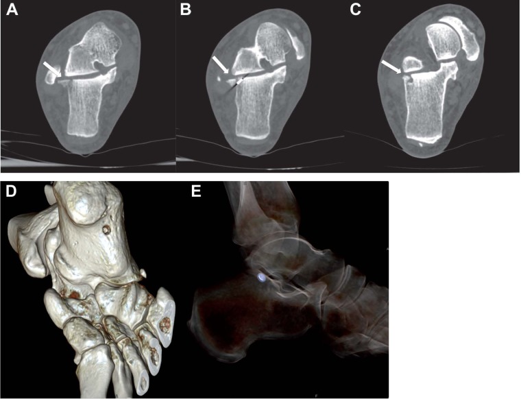
Figure 4.
CT images in axial plane and VR reconstructions show an intra-articular fracture due to bone perforation by a foreign metallic body (A, B and C) (white arrows). VR reconstructions show the inlet hole of the foreign body, with course inside the calcaneal body and the foreign body located near the posterior articular facet (D and E)

