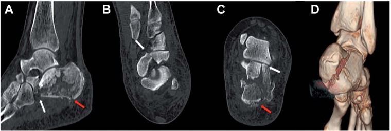
Figure 5.
CT images with MPR in sagittal (A) para-axial (B) and para-coronal planes (C) show intra-articular line fracture in correspondence of the central portion of the posterior facet (white arrows). The excessive impact force generated additional extra-articular fracture lines in correspondence of the posterior heel (red arrows). VR reconstruction, posterior view, better shows extra-articular fractures (D)

