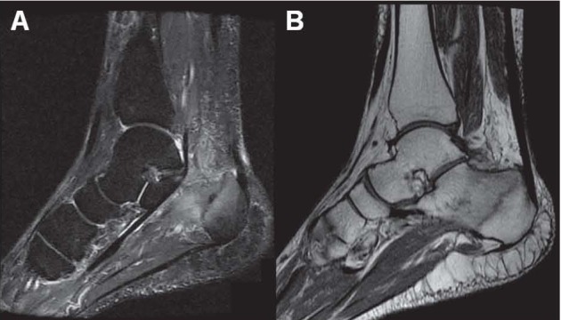
Figure 8.

MRI: sagittal image with STIR (Short TI Inversion Recovery) sequence (A) shows a hypointense fracture line, and a diffuse bone marrow edemigenic imbibition of the calcaneal body and anterior portion of Kager’s triangle, which appears hyperintense. Sagittal image obtained with T1-weighted sequence (B) shows the hypointense fracture line and the hypointense edemigenic imbibition
