Figure 1.

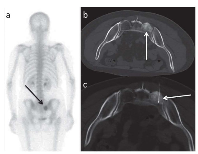
Osteosclerotic lesion of the sacrum. a. and b. Scintigraphy and CT that detected the lesion (arrows); c. image during the treatment: RFA needle inside the lesion (arrow)

Osteosclerotic lesion of the sacrum. a. and b. Scintigraphy and CT that detected the lesion (arrows); c. image during the treatment: RFA needle inside the lesion (arrow)