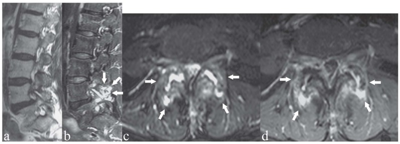
Figure 2.
Patient with bilateral low back pain. a) Sagittal T2-weighted image; b, c) sagittal and axial T2-weighted images with Fat Saturation; d) axial T1-weighted image with Fat Saturation after contrast medium administration. Bilateral facet joint osteoarthritis and synovitis at L4/L5 (arrows). Hyperintensity on T2-weighted images and contrast enhancement of the bone marrow of posterior spinal facet joint articular processes (osteoarthritis) and within the facet joint space (synovitis); the same articular space is enlarged (c). Note the differences between a) and b): “standard” T2-weighted image without fat saturation (a) fails to show the above mentioned pathologic findings that are clearly showed on T2-weighted image with fat saturation (b, c)

