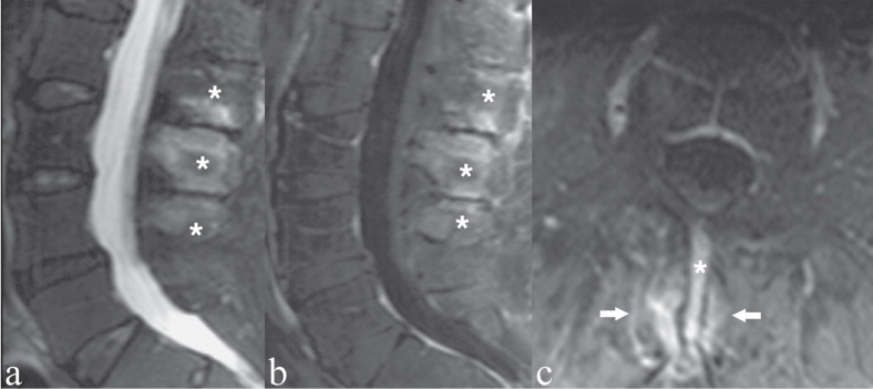
Figure 6.
Patient with persistent low back pain; physical examination showed limited lumbar motion, exacerbation of pain by hyperextension and tenderness on palpation of the interspinous spaces. a) Sagittal T2-weighted image with Fat Saturation; b, c) Sagittal and axial T1-weigthed images with Fat Saturation following contrast medium administration. Lumbar hyperlordosis is present, associated with collision of the spinous processes at L2-L3-L4 (i.e. Baastrup’s phenomenon). The bone marrow of the above-mentioned opposing spinous processes (asterisks) reveals hyperintense areas on T2-weighted image (a) and contrast enhancement (b, c); these findings represent stress-related bony reactive-degenerative changes. The corresponding L2/L3 and L3/L4 interspinous spaces are collapsed. The adjacent muscular fibers (i.e., interspinalis muscles) show contrast enhancement, indicating degenerative-inflammatory muscular alteration (arrows)

