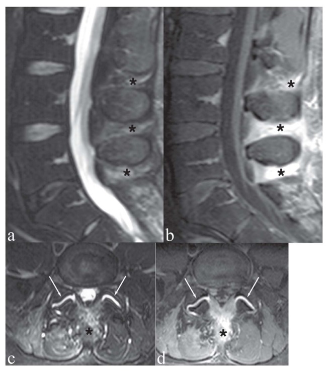
Figure 7.

Patient with low back pain and focal tenderness on the midline. a, c) Sagittal and axial T2-weighted images with Fat Saturation; b, d) Sagittal and axial T1-weighted image with Fat Saturation following contrast medium administration. The interspinous ligaments at L3/L4, L4/L5 and L5/S1 (asterisks) show hyperintensity on T2-weighted images (a, c) and marked contrast enhancement (b, d). These findings indicate degenerative-inflammatory ligamentous changes. Note also bilateral facet joint synovitis (c, d, arrows)
