
Figure 8.
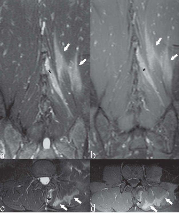
Patient with left-sided low back pain. There was a precise correlation between the MRI findings and the patient’s focus of low back pain. a, c) Coronal and axial T2-weighted images with Fat Saturation; b, d) coronal and axial T1-weighted images with Fat Saturation following contrast medium administration. Hyperintensity on T2-weighted images (a, c) and contrast enhancement (b, d) within the perispinal muscle fibers on the left side: multifidus muscle (asterisk), longissimus and iliocostalis muscles (arrows)