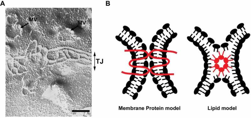
Figure 2.

(A) A freeze fracture image showing tight junction strands in epithelial cells. MV: microvilli, TJ: tight junction. Scale bar, 200 nm. (B) Two models of tight junction. It is widely accepted that tight junctions are formed by the membrane protein, claudin (membrane protein model). The possibility that claudins or other tight junction membrane proteins help assembly and stabilization of a lipid-based strand structure is not completely denied (lipid model).
