
Fig. 2.
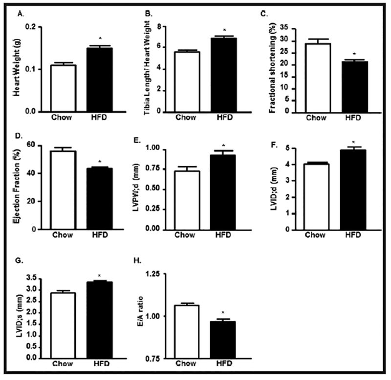
HFD-mediated T2D leads ventricular hypertrophy and induced cardiac dysfunction. (A) Heart weight (HW), (B) Heart weight/Tibia length (HW/TL) ratio in chow and HFD mice. Echocardiographic analysis of (C) left ventricular (LV) fractional shortening (FS), (D) LV Ejection Fraction (EF), (E) Left ventricular posterior wall end diastole (LVPW;d), (F) left ventricle internal diameter, diastole (LVID;d), (G) left ventricle internal diameter, systole (LVID;s) and (H) E/A ratio in control and HFD treated mice (*p<0.05 vs chow).