
Fig. 8.
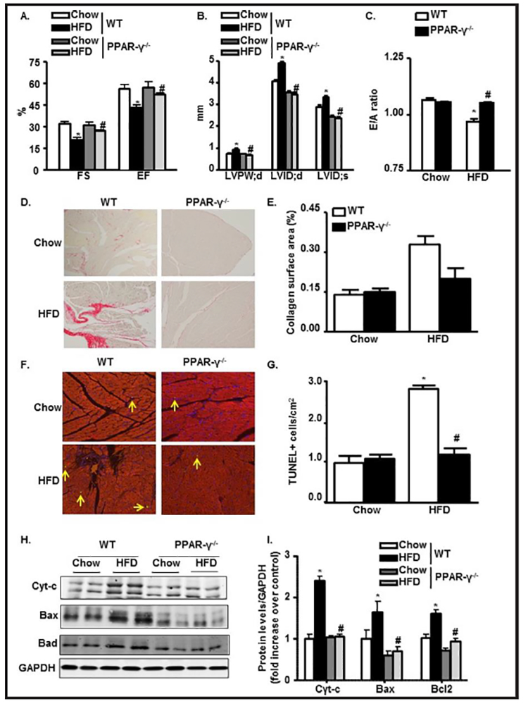
PPAR-γ deletion restores HFD-mediated changes in cardiac function and prevents apoptosis. (A-C) Echocardiographic data showing LVFS, LVEF, LVPWd, LVIDd, LVIDs, and E/A ratio in M-mode in WT and PPAR-γ−/− mice fed with chow or HFD. (D) Collagen deposition with Picrosirius red (200x) in paraffin embedded LV heart in WT and PPAR-γ−/− mice fed with chow or HFD. (E) Semiquantitative analysis of collagen deposition was done by a computer imaging system. (F & G) Apoptotic TUNEL staining (200x) from LV heart and its quantification in WT and PPAR-γ−/− mice fed with chow or HFD. (H-I) Immunoblot from LV heart showing expressions of apoptotic molecules Cytochrome-c, Bax and Bad followed by quantification normalized with GAPDH in WT and PPAR-γ−/− mice fed with chow or HFD (*p<0.05 chow vs HFD, #p<0.05 WT-HFD vs PPAR−/−−HFD)