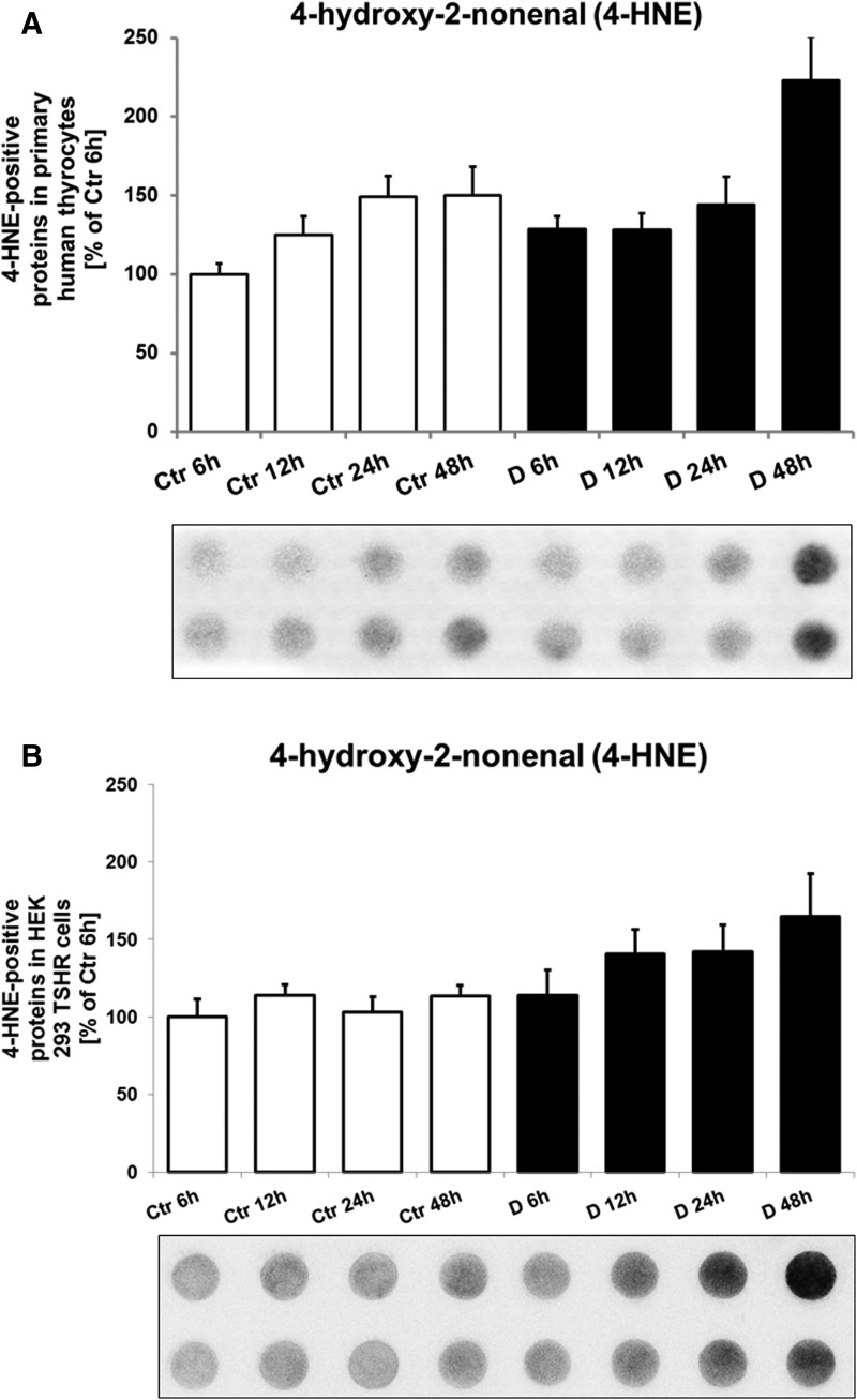
Figure 5.
(A) Representative immunodot-blot of the oxidative stress marker 4-HNE in primary human thyrocyte after incubation with serum from untreated hyperthyroid patients with GD (D for disease, n = 8) and euthyroid healthy control subjects (Ctr, n = 8). The cells were incubated with serum in a time series for 6, 12, 24, and 48 h. In primary human thyrocytes, the 4-HNE marker was significantly higher in the GD 48-h group in comparison with all other groups. The 4-HNE levels were higher in patients with GD vs control subjects at 6 and 48 h (P = 0.02 and P = 0.04, respectively) and in patients with GD at 48-h incubation vs 12 h (P = 0.02) and 6 h (P = 0.01). For Ctr and GD groups, a time-dependent increase was observed. (B) Representative immunodot-blot of the oxidative stress marker 4-HNE in HEK-293 TSHR cells after incubation with serum from untreated hyperthyroid patients with GD (n = 8) and euthyroid healthy control subjects (n = 8). The cells were incubated with serum in a time series for 6, 12, 24, and 48 h. Regarding the HEK-293 TSHR cells, 4-HNE was markedly higher in GD at 48 h vs control subjects at 6 and 48 h (both P < 0.05).

