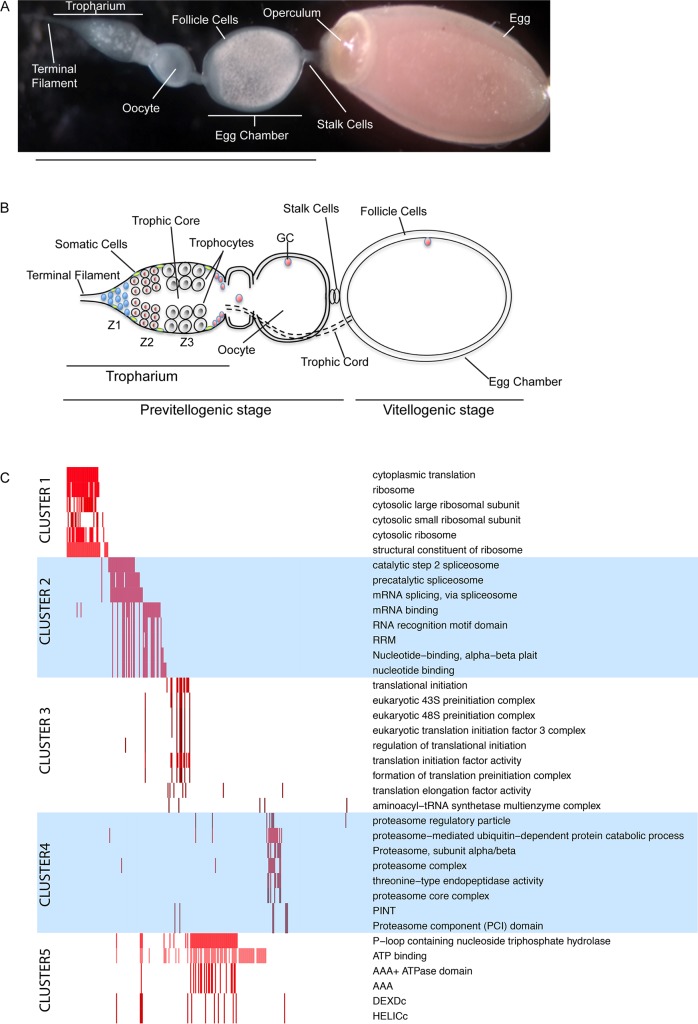
Fig 1. Transcriptomic analyses in early stages of Rhodnius prolixus’ oogenesis.
A) Structure of the ovariole showing the tropharium at the very anterior region, egg chambers in various stages of development and a mature chorionated egg at the very posterior end. The operculum defines the anterior pole of the egg. B) Schematic of the ovariole. The tropharium hosts mitotically active cells in zone 1 at the anterior region and the polyploid trophocytes (i.e. nurse cells) in zone 2 and 3. Oocytes arrested in Meiosis I are located at the posterior region of the tropharium. Each oocyte becomes encapsulated by follicle cells to form the budding egg chamber. Egg chambers in Rhodnius remain connected to the tropharium through the trophic cords, which ensure that nutrients and RNAs produced by the trophocytes are transferred to the growing oocytes. Z1, Zone1, Z2, Zone2, Z3, Zone3, GC, germinal vesicle. C) Functional Analysis of the previtellogenic transcriptome: Five major functional clusters (left) are identified for the 1,050 most expressed genes with high confident homologues in D. melanogaster, obtained by grouping statistically overrepresented terms (right) by their co-occurrence in gene products (x-axis). The matrix in the middle panel visualizes by colors the significance of the p-value computed for a gene.

