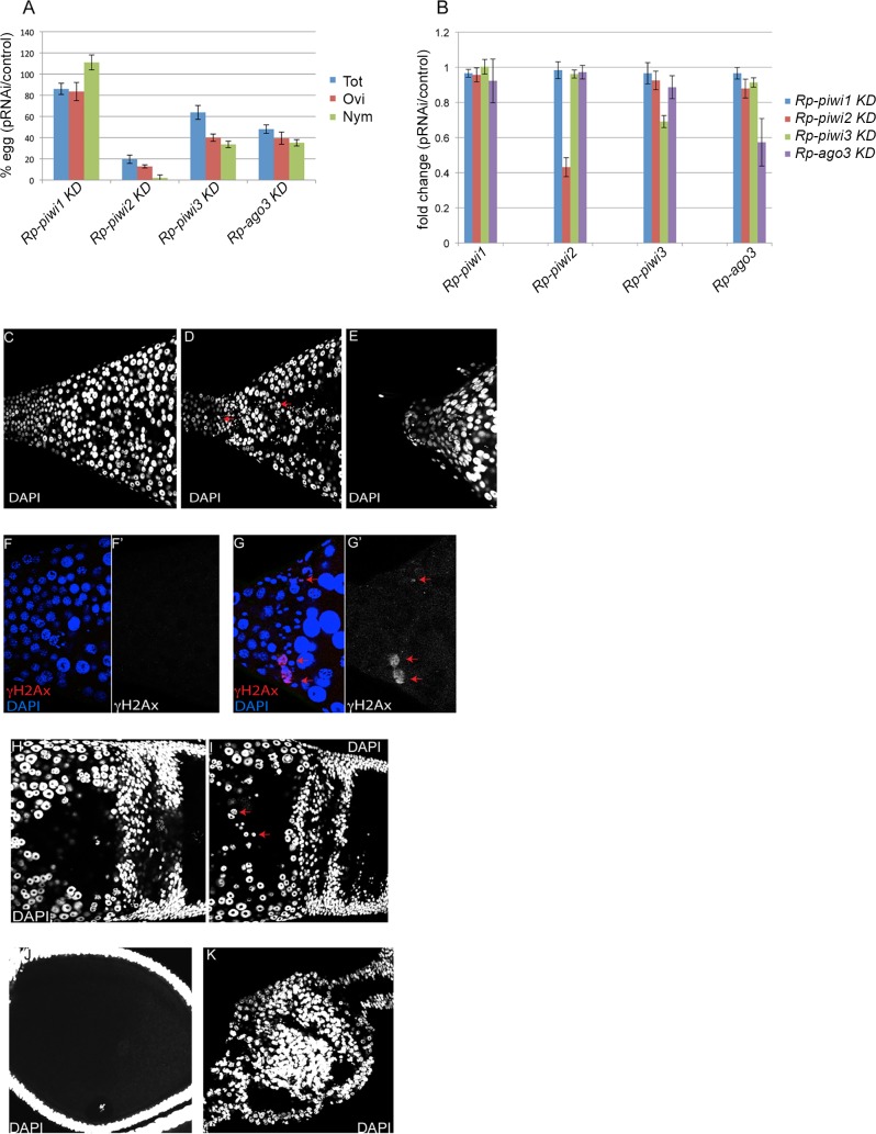
Fig 5. Functional analysis of the Rp-piwi genes.
A) Parental RNAi against the piwi orthologs. Oogenesis and fertility were investigated by dividing the eggs in three groups: total number of eggs (Tot), oviposited eggs (Ovi) and hatched eggs giving rise to first-instar nymphs (Nym). Y-axis displays the percentage eggs produced by Rp-piwi pRNAi treated females compared to control pRNAi females. B) qRT-PCR assays to investigate the expression levels of the Rp-piwi genes after pRNAi treatment. The Y-axis displays the fold difference in the expression levels of the Rp-piwi1, Rp-piwi2, Rp-piwi3 and Rp-ago3 in each of the Rp-piwi KD versus control-injected ovaries. Error bars indicate standard deviation. C-E) Ovarian phenotype of Rp-piwi2 pRNAi females. Zone1 of control (C) and Rp-piwi2 pRNAi (D) ovaries. pRNAi against the Rp-piwi2 gene leads to a reduction in the number of trophocytes and abundant DAPI-positive nuclear debris (red arrows). (E) Zone 1 of Rp-piwi2 pRNAi ovaries is frequently atrophic. F-G) Immunostaining with anti-γH2Ax antibodies (red) and DAPI (blue) in Zone1 of control (F) and Rp-piwi2 pRNAi (G) ovaries. Notice the degenerating nuclei and DAPI-positive particles (red arrow). Single channels for the γH2Ax signal in control (F’) and pRNAi (G’) are shown. H-I) Zone3 and previtellogenic egg chambers of control (H) and Rp-piwi2 pRNAi (I) ovaries stained with DAPI. Degenerating nuclei appear to accumulate in the trophic core of Rp-piwi2 KD tropharia (red arrows). J-K) Control and Rp-piwi2 pRNAi egg chambers. Notice the cortical position of the germinal vesicle and the organized layer of follicle cells surrounding the oocyte in the control (J) ovaries, while egg chambers appear collapsed or atrophic upon pRNAi for Rp-piwi2 (K).

