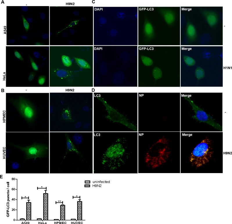
Fig 1. Autophagsomes were formed and accumulated after IAV infection in various cell types including endothelial cells.
The cells were transiently transfected with plasmids expressing GFP-LC3 and thereafter infected with IAV H9N2 or H1N1. The cells were observed for GFP-LC3 distribution in the cytoplasm by confocal fluorescence microscopy at 24 hrs post infection. DAPI was used for nuclear staining. Scale bar, 20μm. (A) A549 and HeLa cells infected with H9N2; (B) HPMECs and HUVECs infected with H9N2; (C) HeLa cells infected with an H1N1 IAV; (D) The presence of viral NP and LC3 in H9N2-infected HPMECs. Scale bar, 20μm; (E). Quantitative analyses of autophagosomes in infected and non-infected cells of various types (** p<0.05; ***p<0.01).

