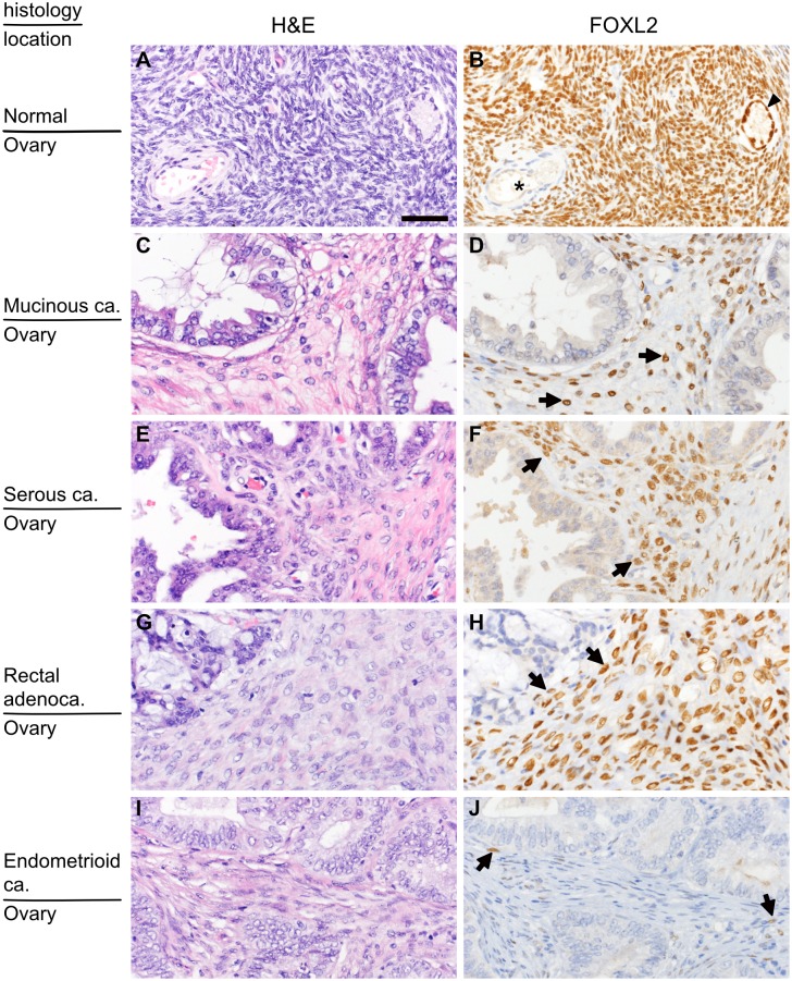
Fig 1. Distribution of FOXL2-positive stromal cells in the ovary.
(A and B) Normal ovarian cortex. Almost all stromal cells as well as granulosa cells (arrow head) show nuclear positivity for FOXL2. By contrast, none of the cells that constitute blood vessels (asterisk) have nuclear FOXL2. (C and D) Primary mucinous carcinoma. (E and F) Primary serous carcinoma. (G and H) Secondary ovarian tumor (ovarian metastasis of rectal adenocarcinoma). Most stromal spindle cells show nuclear positivity for FOXL2. (I and J) Primary endometrioid carcinoma, a rare example of primary ovarian cancer containing only a few FOXL2-postive cells. H&E (left panels) and FOLX2 immunostaining of the corresponding area (right panels; only nuclear staining is considered positive). Arrows indicate FOXL2-positive cells. The bar in (A) indicates 50μm, and the magnification is identical for all the pictures.

