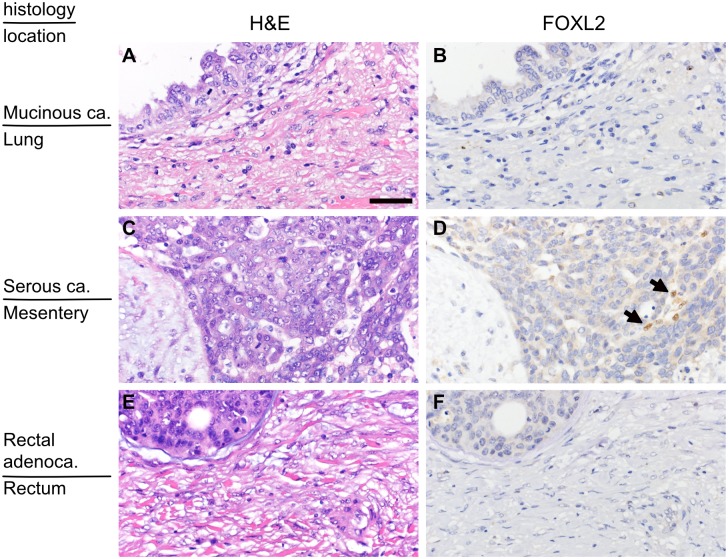
Fig 2. Distribution of FOXL2-positive stromal cells in extraovarian lesions.
(A and B) A metastatic site of mucinous ovarian carcinoma in the lung (the same case as Fig 1C and 1D). There are no FOXL2-positive stromal cells. (C and D) A peritoneal seeding site of serous ovarian carcinoma (the same case as Fig 1E and 1F). There are few FOXL2-positive cells. (E and F) The primary site of rectal cancer that caused secondary ovarian tumor (the same case with Fig 1G and 1H). There are no FOXL2-positive stromal cells. H&E (left panels) and FOLX2 immunostaining of the corresponding area (right panels). Arrows indicate FOXL2-positive cells. The bar in (A) indicates 50μm, and the magnification is identical for all the pictures.

