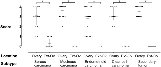
Fig 3. The proportion of FOXL2-positive cells in cancer stroma.
The proportion of FOXL2-positive cells on each tissue section was scored using 5-tired scale and the results were plotted. High percentages of cancer stromal cells were FOXL2-positive in the ovary, whereas there were almost no FOXL2-positive cells outside the ovary (Ext-O). Asterisks indicate a significant difference between the groups by Mann-Whitney test (p < 0.05).

