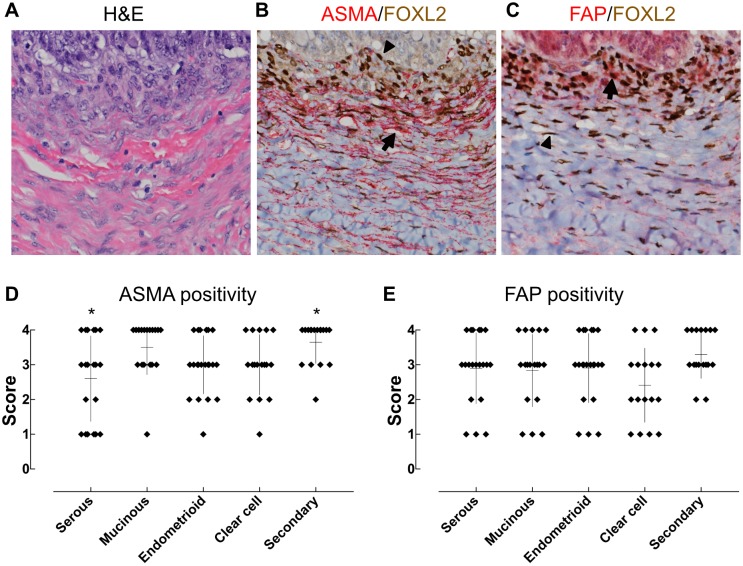
Fig 4. Expression of ASMA and FAP by FOXL2-positive ovarian cells.
(A-C) Tumor tissues were double stained for FOXL2 and ASMA or FAP. Nuclear FOXL2 was stained brown whereas cytoplasmic ASMA and FAP were stained red in B and C, respectively. Some FOXL2-positive cells were also stained positive for ASMA or FAP (arrows), but others were negative (arrow heads). (D and E) The frequency of ASMA and FAP positive cells in FOXL2-positive cells. ASMA and FAP were variously but at least focally expressed by FOXL2-positive cells of all cases. Asterisks indicate a significant difference between two groups (Kruskal-Wallis test followed by multiple comparison).

