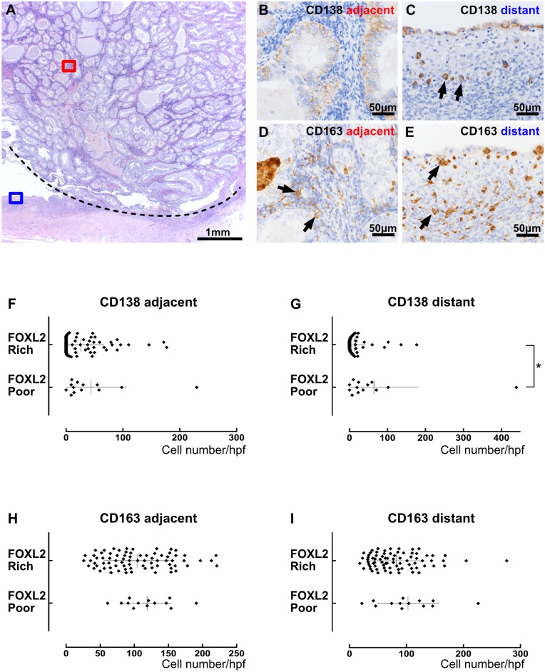
Fig 5. Correlation between chronic inflammation and FOXL2-positive cells in ovarian cancer stroma.
(A-E) An example of endometrioid carcinoma where FOXL2-positive stromal cells were scant (FOXL2-poor). (A) Low magnification of H&E. The area above the dotted line is cancer tissue and the area below the line is a pre-existing endometrial cyst. (B and C) CD138 immunostaining. Tumor area (B: red square in A) contained no plasma cells and nontumor area (C: blue square in A) had a few plasma cells (arrows). (D and E) CD163 immunostaining. Both tumor area (D) and nontumor area (E) had some macrophages (arrows). (F-I) The number of CD138-positive plasma cells (F and G) or CD163-positive macrophages (H and I) in one high power field was plotted, comparing between cases with FOXL2-poor stroma (score1 and 2) and those with FOXL2-rich stroma (score 3 and 4). The presence of plasma cells apart from tumor cells correlated with FOXL2-positive cell population, whereas the presence of plasma cells near tumor cells or macrophages in any place did not. The asterisk indicates a significant difference between the groups by Mann-Whitney test (p < 0.05).

