
Extended Data Figure 4. EM analysis of cell types in the DG of a 13 year old and adult human brain; absence of SGZ precursor cells or immature neurons.
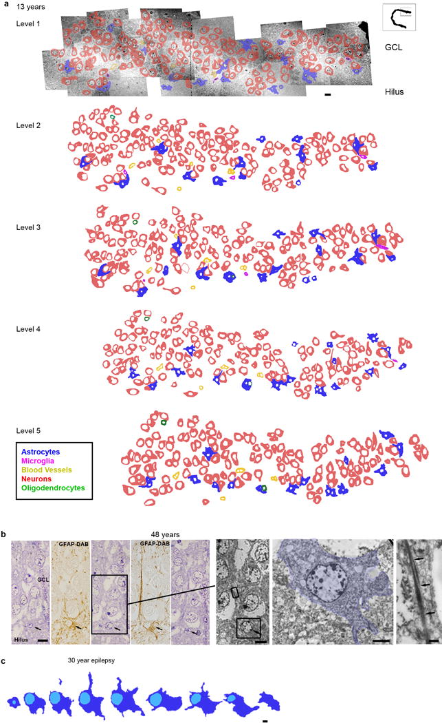
a, Reconstruction of 5 ultrathin sections (separated by 1.5 μm) from the 13 year old GCL with outlines of cell membranes. Colors corresponding to the different cell types defined by their ultrastructural characteristics are indicated in the key. No clusters or isolated cells with young neuronal ultrastructure were found. Cells associated in small groups were identified as astrocytes, oligodendrocytes or microglia. b–c, reconstructions of astroglial cells next to the GCL searching for possible examples of RA in the adult human DG. b, example of an astrocyte with radial morphology in the adult GCL. Five serial semithin sections of this astrocyte (black arrows) next to the GCL of a 48 year old DG ; alternating semithin sections show that this cell is GFAP+. This cell extends a thin radial fiber through the GCL, but has multiple processes (stellate morphology) in the hilus. Boxed area shows the ultrastructure from the indicated semi-thin section of this astrocyte (pseudo-coloured in blue) and the bundles of intermediate filaments present in the expansion (arrows). c, Another example of a serially reconstructed astrocyte in the DG of a 30 year old epileptic case (separated by 1.4 μm) showing a short radial expansion and processes into the hilus. Scale bars: 10 μm (a, b semithin and TEM), 5 μm (c), 2 μm (b, soma), 500 nm (b, intermediate filaments).