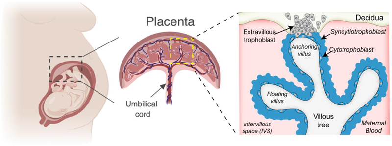
Figure 2: Human placental Structure.
A zoomed in villous tree shows floating and anchoring villi. The villous trees are lined by syncytiotrophoblasts and an inner layer of cytotrophoblasts (that become more discontinuous throughout pregnancy) that fuse to replenish the outer syncytial layer. Invasive extravillous trophoblasts extend from the villous tree into the maternal decidua and both anchor the placenta to the uterine wall and remodel the maternal microvasculature.

