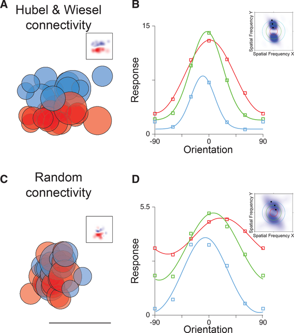
Figure 1. Receptive Fields, Random Connectivity, Spatial Frequency (SF) Tuning, and Orientation Tuning.
(A) Hubel and Wiesel connectivity in which ON (red) and OFF (blue) thalamo-cortical afferents, with spatial receptive fields indicated by each circle, converge onto a neuron in primary visual cortex. The summation of these afferent receptive fields generates a Gabor-like receptive field in visual cortex (inset).
(B) Orientation preference does not change with SF for such receptive fields. Tuning curves of the temporal modulation of the response for low (red), medium (green), and high (blue) spatial frequencies are plotted. In frequency space, these receptive fields maintain a peak response at a consistent angle that points toward the origin at the midpoint of the graph (inset).
(C) Random connectivity from the lateral geniculate nucleus (LGN) in which ON and OFF thalamo-cortical neurons with similar spatial receptive fields converge on cortical neurons also generates orientation selectivity in the temporal modulation of the response. The linear summation of LGN ON and OFF neuron receptive fields shows oriented profiles (inset). Scale bar indicates 35 degrees.
(D) Orientation preference shifts for random connectivity as SF changes. Orientation tuning curves are plotted as in (B). In frequency space, these receptive fields tilt in a manner that does not project back to the origin.

