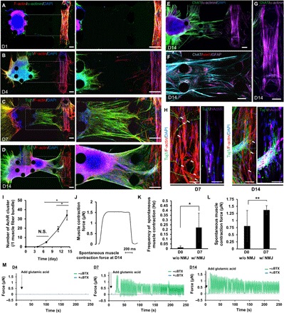
Fig. 3. Neural outgrowth and formation of a human motor unit along with NMJ.

(A) Injection of an MN spheroid into the left compartment of the microfluidic device with collagen gel on D0. (B) MNs started neural outgrowth toward the muscle fiber bundle on D4. (C) After 7 days of coculture, many axons reached the muscle fiber bundle and end feet of neurons attached to myotubes, resulting in the formation of NMJs. (D) By D4 of coculture, thick neural fibers were observed. Soma of MNs also migrated from the original position. (E) Characterization of mature MNs stained by ChAT and islet1 (F) and a mature myotube (G) after D14. Scale bars, 100 μm (A to G). (H) Localization of nAChR on the muscle fiber bundle on D7 and D14. Scale bars, 10 μm. (I) The number of clusters of nAChRs on single muscle fiber bundles increased over time. n = 4. (J) Muscle contraction force estimation by pillar displacement on D14 of coculture. (K) Frequency of spontaneous muscle contraction and spontaneous muscle contraction force (L) on D0 (before NMJ formation, without NMJ) and D7 (after NMJ formation, with NMJ) without glutamic acid stimulation. n = 6. (M) Measurement of muscle contraction force by adding glutamic acid on D4, D7, and D14. **P < 0.05; *P < 0.01, Student’s t test and one-way ANOVA. Error bars ± SD.
