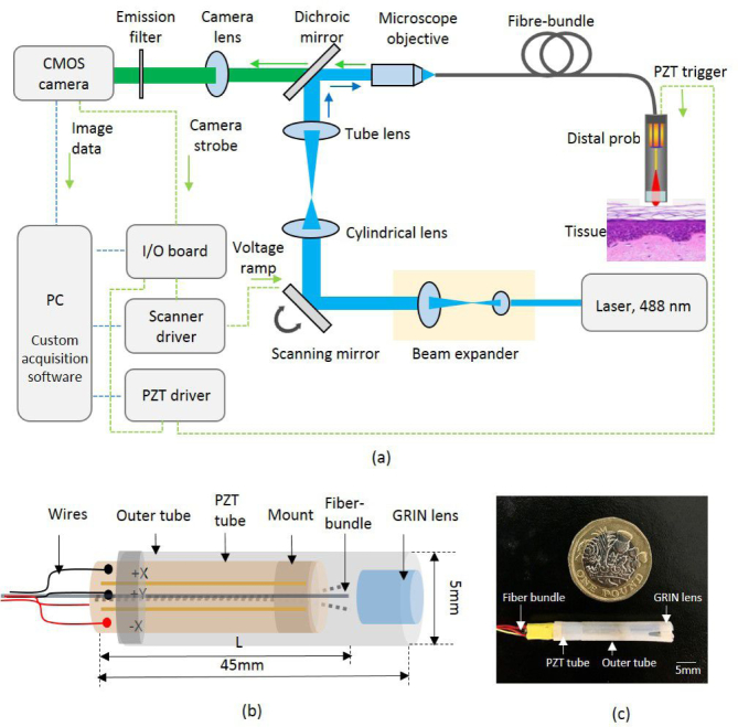
Fig. 1.
Schematics of (a) the line-scan confocal laser endomicroscopy (LS-CLE) system with (b) fiber-shifting distal probe. The proximal face of the fiber bundle is placed at the focal plane of the LS-CLE and the distal end is actuated by a PZT tube behind a GRIN lens with 1.92X magnification. (c) A photograph of the assembled 3D printed probe holder tube with 5 mm outer diameter. A UK one pound coin is shown for scale.

