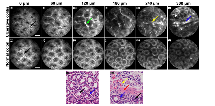
Fig. 11.
Different depth images of tissues of normal colon and colon with ulcerative colitis. (a-f) Different depth images of ulcerative colitis. (g-l) Different depths images of normal tissues of colon. (a) and (g) Columnar epithelial cells (black arrows) and crypts can be seen on the surface of both tissues. (c) Ulcerative colitis shows ICG leakage (green arrow) between crypts. (d) Ulcerative colitis shows irregular crypts with ICG leakage. (e) Blood vessel (yellow arrow) can be seen without crypt structures. (f) Fibers with strong fluorescence in layer of muscularis mucosae can be seen in ulcerative colitis (white arrow and blue arrow). (g-l) Signal intensity falls and there is hardly a change in the structure with increasing imaging penetration depth. (m) and (n) correspond to histologic specimens of normal and ulcerative colitis tissues. In (m) and (n), black arrows: crypts; blue arrows: columnar epithelial cells; yellow arrow: blood vessel; red arrow: fiber in muscularis mucosae. Scale bars: 50 μm.

