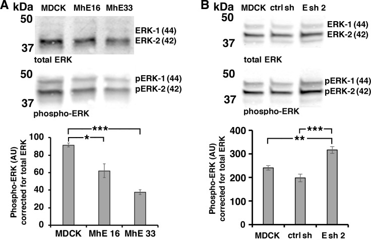
Fig 3. EpCAM inhibits ERK activation.
(A) Wildtype MDCK, and MDCK lines overexpressing human EpCAM (MhE16, MhE33) were SDS extracted one day after plating at subconfluent cell density; levels of ERK and phospho-ERK were analyzed in the same immunoblot. The graph shows quantification of combined 44 and 42 kDa phospho-ERK protein levels normalized to combined 44 and 42 kDa ERK protein levels in the same sample. Error bars: S.E.M. of three independent samples for each cell line; p values derived from unpaired Student’s t test: * p = 0.023 for MhE16 to MDCK and *** p = 0.0001 for MhE33 to MDCK. (B) Phospho-ERK levels in MDCK cells, control shRNA-expressing MDCK cells (ctrl sh) and EpCAM-depleted MDCK cells (Esh2) were analyzed as in (A). Error bars: S.E.M. of six independent samples for each cell line; p values derived from unpaired Student’s t test: ** p = 0.0013 for Esh2 to MDCK and *** p = 0.0003 for Esh2 to ctrl sh.

