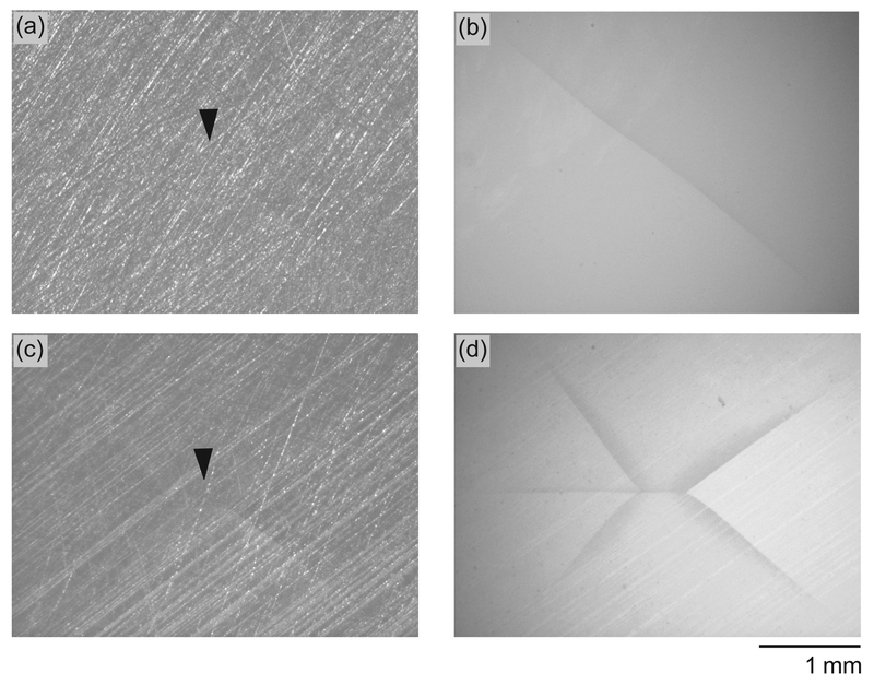
Figure 3.
Representative stereo optical microscopy images show surface features of (a) 3Y-TZP and (c) 5Y-PSZ discs fractured at 1010 N and 774 N, respectively. Images were obtained using reflected light illumination. Arrows indicate the contact area, demonstrating the absence of near-contact induced top surface cone cracks. (b) and (d) are images of the same specimens, but acquired using transmitted light illumination to reveal the far-field flexural induced radial cracks initiated at the cementation surface of the ceramic discs. Images were taken on ceramic discs that have been “peeled off” of their dentin analog composite substrate after the Hertzian load-to-fracture test.

