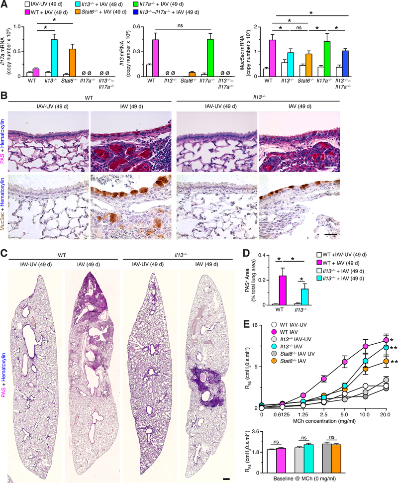
FIGURE 8.
Persistence of IL-13–STAT6-linked chronic lung disease after IAV infection. (A) Lung levels of Il17a, Il13, and Muc5ac mRNA in WT, Il13–/–, Stat6–/–, Il17a–/–, and Il13–/––Il17a–/– mice at 49 d after infection with IAV (2 pfu) or IA-UV. (B) Representative PAS-hematoxylin staining and Muc5ac-hematoxylin immunostaining of lung sections from WT and Il13–/–mice at 49 d after infection with IAV (2 pfu) or IAV-UV. Bars=400 μm. (C) PAS-hematoxylin staining of lung sections from WT and Il13–/–mice at 21 d after infection with IAV (2 pfu) or IAV-UV. Bar=1 mm. (D) Quantitation of PAS-positive areas in images in (D). (E) Levels of airway reactivity using response RRS to inhaled MCh and for baseline RRS for conditions in (A). For (A)-(E), values are representative of 3 separate experiments (n≥8 mice per condition in each experiment). For (A), (C), and (D), * indicates p<0.05.

