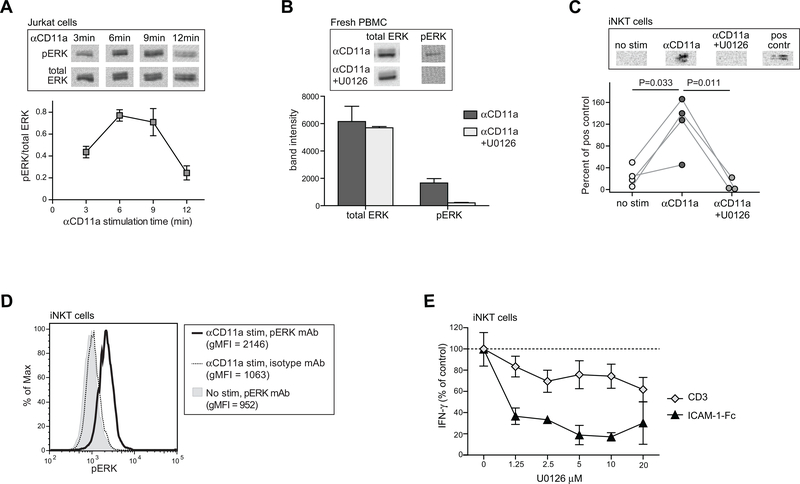
Figure 4. iNKT cell IFN-γ secretion in response to LFA-1 stimulation is dependent on ERK phosphorylation.
A) Jurkat T cells were stimulated with anti-CD11a mAb for the indicated times, then lysed and subjected to Western blotting using detection antibodies against either total ERK or phospho-ERK. Images at the top show the phospho-ERK and corresponding total ERK results from a representative Western blot analysis; the plot below shows the means and standard deviations of the phospho-ERK signal normalized by the corresponding total ERK signal from three replicate Western blot analyses. B) Freshly isolated PBMCs were stimulated for 5 min with anti-CD11a in the presence or absence of 5 μM of the MEK inhibitor U0126. Images at the top show phospho-ERK and corresponding total ERK Western blot results. The plot below shows the band intensity (mean, std dev)from 3 independent analyses. C) Cultured iNKT cells (FoB short-term polyclonal line) were stimulated with anti-CD11a mAb for 5 minutes in the presence or absence of 5 μM U0126 and lysates were Western blotted to detect phospho-ERK. Positive control indicates Jurkat cells that were stimulated with 20nM 12-O-Tetradecanoyl-phorbol-13-acetate for 10 minutes. Plot below shows the aggregated results from 4 independent analyses, with band intensities for iNKT cell p-ERK normalized by the signal from the corresponding positive control band run in parallel. D) iNKT cells (318D short-term polyclonal line) were stimulated with anti-CD11a mAb for 5 minutes or mock-treated, then fixed and permeabilized, stained using an antibody against p-ERK or an isotype-matched negative control, and analyzed by flow cytometry. E) iNKT cells (FoB short-term polyclonal line) were incubated for 24h with plate-bound ICAM-1-Fc or with soluble anti-CD3 mAb, in the presence of the indicated concentrations of U0126, and secreted IFN-γ was quantitated by ELISA. The plot shows the amount of IFN-γ as a percentage of the amount produced in each condition in the absence of U0126; symbols represent the means and standard deviations (not always visible on the scale shown) of four replicates. Similar results were obtained in two additional independent analyses using the FoB polyclonal and PP1.3 clonal iNKT cell lines.

