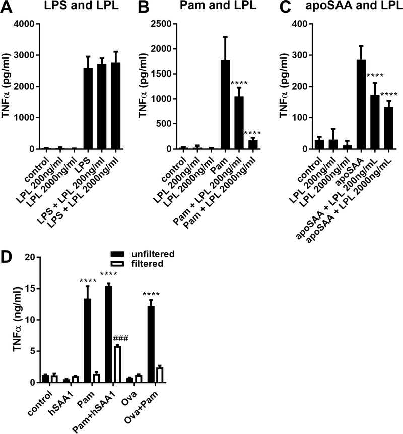
Figure 5.
Lipopeptides associated with SAA confer its ability to stimulate TNFα production. 100 ng/ml LPS (A), 100 ng/ml Pam3CSK4 (Pam, B), and 1¼g/ml apoSAA (C) were incubated with lipoprotein lipase (LPL), and then used to stimulate J774 cells. Culture supernatants were collected 24 hours later and TNFα concentrations were measured by ELISA. Human SAA1 from Origene produced in HEK cells (hSAA1) or ovalbumin (Ova) was unexposed or exposed to Pam3CSK4 (Pam) overnight (control), and some of the preparation was subjected to concentration of proteins >3 kDa using concentrated using Amicon Ultra centrifugal concentrators (filtered). Control and filtered preparations were used to stimulate J774 cells for 24 hours after which TNFα concentrations in culture media were measured by ELISA (D). Data are mean ± SEM and are representative of three (A-C) or two (D) independent experiments with n = 4 per group. Statistics were analyzed using one-way ANOVA with Dunnett’s test for multiple comparisons (A-C) or two-way ANOVA with Tukey’s test for multiple comparisons (D). **** p < 0.0001 compared to Pam (B), apoSAA (C), or control (D). ### p < 0.001 compared to unfiltered Pam+hSAA1 (D).

