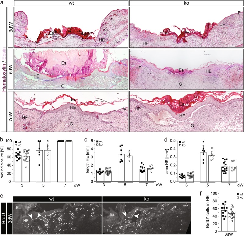
Fig. 2.
Nrf3 is dispensable for wound healing. a H/E staining of sections from 3-day, 5-day, and 7-day wounds of 8–9-week-old mice. E: Epidermis; Es: Eschar; G: Granulation tissue; HE: Hyperproliferative wound epidermis; HF: Hair follicle. b–d Morphometric analysis of (b) percentage of wound closure, (c) length HE, and (d) area HE of 3-day, 5-day, and 7-day wounds. e, f Immunofluorescence staining for BrdU with arrowheads pointing to representative BrdU-positive cells (e) and quantitative analysis (f) of BrdU-positive cells in the HE of 3-day wounds. Bars: 500 μm (a) and 200 μm (f). Scatter plots show mean and SD in b–d and f. Each data point represents the result from an individual wound

