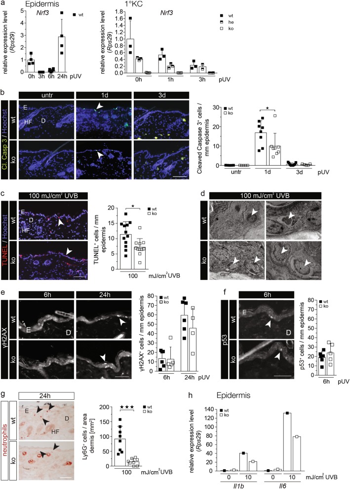
Fig. 3.
Loss-of-Nrf3 protects keratinocytes from UVB-induced apoptosis in vivo. a RNA samples from epidermis of mice prior to and post irradiation with 100 mJ/cm2 UVB (a) or from primary keratinocytes of wt and heterozygous or homozygous Nrf3-ko mice prior to and at different time points after irradiation with 10 mJ/cm2 UVB (b) were analyzed by qRT-PCR for expression of Nrf3 relative to Rps29. b–c: Cleaved caspase 3 (b) or TUNEL staining (c; color changed from green to red) using sections from untreated (untr) skin or 1 d or 3 d after irradiation with 100 mJ/cm2 UVB. Nuclei were counterstained with Hoechst (blue). Bar (b, c): 100 µm. Positive cells per length epidermis are shown in the scatter plots. d Electron microscopy of UVB-irradiated epidermis. Upper left: Basal keratinocyte (arrowhead) that has lost cell–cell and cell-matrix contacts and reveals a hyperdense cytoplasm and a condensed nucleus as a sign of early apoptosis. Upper right: Debris of a keratinocyte (arrowhead) at a later stage of apoptosis with complete detachment from the basement membrane. Lower left: Basal keratinocytes (arrowheads) from Nrf3-ko mice show only slight condensation and shrinkage of the nuclei without signs of cytoplasmic alteration. Lower right: Some swollen and hypo-dense keratinocytes with signs of karyolysis (arrowheads) in the basal layer of Nrf3-ko mice, but without detachment from the basement membrane. Bar: 3 µm. e–g Sections from skin 24 h after irradiation with 100 mJ/cm2 UVB were stained with antibodies against γH2AX, p53 or Ly6G, and the numbers of γH2AX- or p53-positive keratinocytes or Ly6G-positive neutrophils per mm epidermis or per area dermis, respectively, were determined. 15–25 microscopic fields of skin sections per mouse were analyzed. Bars: 25 µm (e, f) and 100 µm (g). h RNA from the skin of wt and Nrf3-ko mice prior to and 24 h after UVB irradiation was analyzed by qRT-PCR for Il1b and Il6. RNAs were pooled from 5–6 mice per genotype and treatment group. The result was reproduced with RNAs from an independent experiment with different mice. Arrowheads point to apoptotic keratinocytes (b, c, e) or Ly6G-positive neutrophils (g). E: Epidermis, D: Dermis, HF: Hair follicle. Scatter plots in a–c and e–g show mean and SD. Each data point represents the result from an individual mouse

