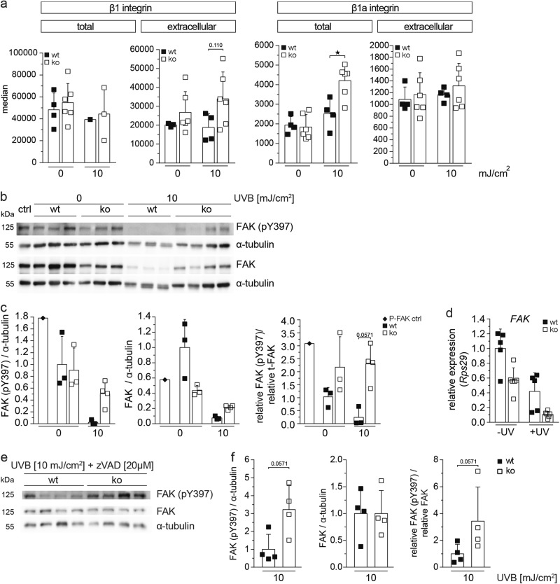
Fig. 8.
Loss-of-Nrf3 enhances adhesion signaling prior to and following UVB irradiation. a Immortalized keratinocytes from wt and Nrf3-ko mice were permeabilized (total) or not (extracellular) and stained with antibodies against the integrin subunits β1 or active integrin β1, either prior to or 24 h after irradiation with 10 mJ/cm2 UVB and analyzed by flow cytometry. b–d Immortalized keratinocytes from wt and Nrf3-ko mice were irradiated with 10 mJ/cm2 UVB and analyzed by western blotting prior to and 24 h post irradiation for phosphorylated and total FAK and for α-tubulin (loading control) (b). The ratios of phosphorylated FAK, respectively total FAK to α-tubulin, as well as phosphorylated to total FAK are depicted in the graphs (c). The P-FAK positive control (P-FAK pos. ctrl) was obtained by incubating keratinocytes in fresh EGF-containing culture medium for 20 min immediately before sampling. Alternatively, cells were analyzed by qRT-PCR for expression of Fak relative to Rps29 (d). e Immortalized keratinocytes were treated with 20 µM zVAD before irradiation with 10 mJ/cm2 UVB and analyzed by western blotting 24 h post irradiation for phosphorylated and total FAK and for α-tubulin. The ratios of pFAK, respectively total FAK to α-tubulin, as well as pFAK to total FAK are depicted in the graphs (f). All results shown are representatives of at least two independent experiments

