Fig. 2.
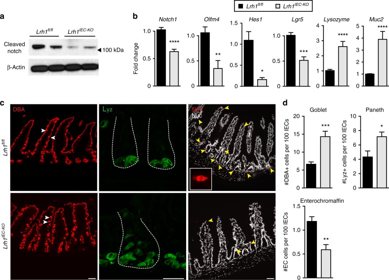
Lrh1IEC-KO mouse organoids have diminished Notch activation. a Representative immunoblot for cleaved Notch1 in Lrh1fl/fl (left two lanes) and Lrh1IEC-KO (right two lanes), n = 4 per condition. b Fold change in expression by RT-qPCR in Lrh1fl/fl (black) and Lrh1IEC-KO (gray) organoids for Notch1 and the Notch target genes Olfm4 and Hes1. Relative expression of the stem cell marker Lgr5, Paneth cell marker Lysozyme, and goblet cell marker Mucin2 are also shown. Minimum of three replicates per condition. c Histology of Lrh1fl/fl and Lrh1IEC-KO small intestine showing goblet (DBA, red, left), Paneth (lysozyme, green, middle) and enterochromaffin cells (5HT, red, right and inset). Scale bar = 50 μm. Intestinal crypts are outlined with dashed white line in middle panel. d Quantitation of epithelial subtype distribution, n = 3 animals per condition. For b and d, error bars are SEM using Student’s t test (unpaired, two tailed) with p values of *p = < 0.05, **p = < 0.01, ***p = < 0.001, and ****p = < 0.0001

