Fig. 1.
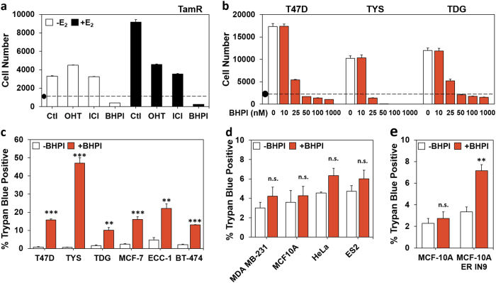
BHPI kills ERα+ breast and endometrial cancer cells. a TamR cell proliferation after 4 days in 8% charcoal:dextran-treated calf (CD-calf) serum with the indicated treatments: E2 100 pM; z-4-hydroxytamoxifen (OHT, the active form of tamoxifen) 1 µM; ICI/fulvestrant 1 µM; BHPI 100 nM. (•) Indicates starting cell number (day 0). b Dose−response study of the effect of increasing concentrations of BHPI on proliferation of T47D, TYS, and TDG cells after 4 days. a, b Alamar Blue assays (n = 8 biological replicate experiments). c Automated trypan blue exclusion assays of ERα-positive cells after 24 h treatment with 1 µM BHPI. d Trypan blue exclusion assays of ERα-negative cells after 24 h treatment with 1 µM BHPI. e Trypan blue exclusion assays of isogenic MCF10A and MCF10AER IN9 cells after 24-h treatment with 1 µM BHPI. c–e Data is mean ± s.e.m. (n = 3 biological replicate experiments). For (c–e) **p < 0.01, ***p < 0.001, n.s. = not significant by Student’s t test

