Fig. 1.

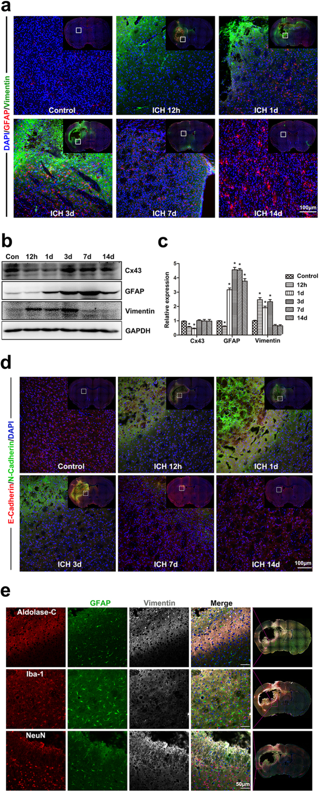
The astroglial-mesenchymal phenotype switching of astrocytes in ICH mouse brain. a Immunofluorescence staining for Vimentin (green) and GFAP (red) in normal and ICH mouse brain at 12 h, 1d, 3d, 7d, and 14d. Bar = 100 μm. Vimentin was undetectable in normal brain, whereas after ICH, it was intensely expressed in the hematoma margin. The hematoma was completely resolved at 7d post-ICH. The normal brain had a baseline expression of GFAP, reactive astrocytes with intensive GFAP expression and hypertrophy of cellular processes were observed in the peri-lesion area at 3d and 7d post-ICH. At 14d post-ICH, glial scar formed with intensive GFAP staining. b Western blotting analysis of Cx43, GFAP and Vimentin expression in normal and ICH mouse brain at 12 h, 1d, 3d, 7d, and 14d. c The results of densitometric analysis of the bands were plotted as mean ± SEM of five independent experiments. Vimentin expression was significantly increased after ICH, reached a peak at 3d and returned to normal at 7d post-ICH. GFAP expression was significantly increased at 1d, 3d, 7d, and 14d post-ICH, reached a peak at 7d post-ICH. There was a transient decrease in GFAP expression at 12 h post-ICH. *p < 0.05, **p < 0.01 compared with control. d Immunofluorescence staining for E-Cadherin (red) and N-Cadherin (green) in normal and ICH mouse brain at 12h, 1d, 3d, 7d, and 14d. Bar = 100 μm. e Immunofluorescence triple staining with Adolase-C + GFAP + Vimentin, Iba-1 + GFAP + Vimentin, NeuN + GFAP + Vimentin in the peri-lesion area of ICH brain. Bar = 50 μm
