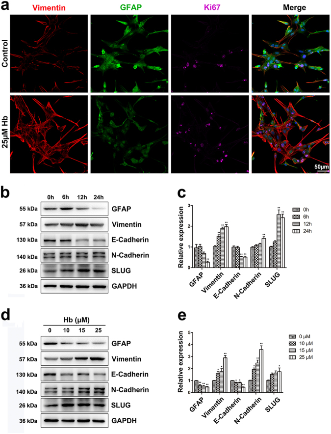
Fig. 3.
Hb induced epithelial-mesenchymal transition in astrocytes. a Immunofluorescence staining of astrocytes for Vimentin (red), GFAP (green), and Ki67 (purple). The cell nuclei were counterstained with DAPI (blue). Bar = 50 µm. b,d Western bloting analysis of GFAP, Vimentin, E-Cadherin, N-Cadherin, and SLUG expression in astrocytes treated with Hb for indicated times and doses. c,e The results of densitometric analysis of the bands were plotted as mean ± SEM of three independent experiments. Hb treatment decreased GFAP and E-Cadherin expression and increased Vimentin, N-Cadherin, and SLUG expression. *p < 0.05, **p < 0.01 compared with control (0 h or 0 µM)

