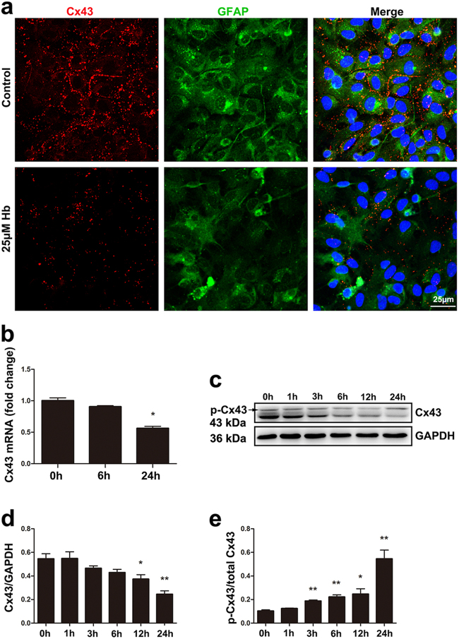
Fig. 4.
Hb exposure induced Cx43 downregulation in astrocytes. a Immunofluorescence staining for connexin 43 (red) and GFAP (green) in astrocytes treated with or without 25 μM Hb. The cell nuclei were counterstained with DAPI (blue). Cx43 was expressed on the cell membrane where astrocytes contact. After Hb treatment, Cx43 expression was decreased. Bar = 25 µm. b Cx43 mRNA expression in control and astrocytes treated with 25 μM Hb for 6 or 24 h. Data were normalized against the internal reference GAPDH. The fold change values were calculated by normalizing to control samples. The results were plotted as mean ± SEM of three independent experiments. Hb treatment for 24 h decreased Cx43 mRNA expression by half. *p < 0.05 compared with control (0 h). c Western blotting analysis of Cx43 expression in astrocytes treated with 25 μM Hb for 0, 1, 3, 6, 12, or 24 h. d,e The results of densitometric analysis of total Cx43 and p-Cx43/total Cx43 were plotted as mean ± SEM of three independent experiments. Hb-treatment decreased Cx43 expression and increased the ratio of p-Cx43/total Cx43. *p < 0.05, **p < 0.01 compared with control (0 h)

