
Fig. 5.
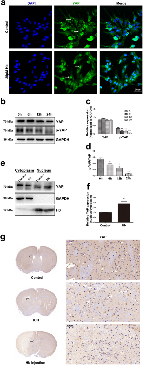
Hb treatment induced YAP nuclear translocation in astrocytes. a Immunofluorescence staining of astrocytes with YAP (green) antibody. The cell nuclei were counterstained with DAPI (blue). The arrows indicate the differential localization of immunopositive products in the nuclei. Hb treatment promoted YAP nuclear translocation. Bar = 25 µm. b Western blotting analysis of YAP and p-YAP expression in astrocytes treated with 25 μM Hb for 0, 6, 12, or 24 h. c,d The results of densitometric analysis of YAP and p-YAP and p-YAP/YAP were plotted as mean ± SEM of three independent experiments. p-YAP and p-YAP/YAP expression was decreased upon Hb exposure. *p < 0.05, **p < 0.01 compared with control (0 h). e Western blotting analysis of cytoplasmic and nucleus extraction samples from control and 25 μM Hb treated astrocytes with YAP antibody. GAPDH and H3 were used as loading control for cytoplasmic and nucleus protein, respectively. f The histogram showing the results of densitometric analysis of nucleus/cytoplasmic YAP expression in control and 25 μM Hb treated astrocytes. The results were normalized to control. Hb treatment significantly increased YAP nuclear expression. *p < 0.05, **p < 0.01 compared with control. g Immunohistochemistry staining of YAP in control brain and the peri-lesion area of ICH model and Hb-injection model. Bar = 50 μm