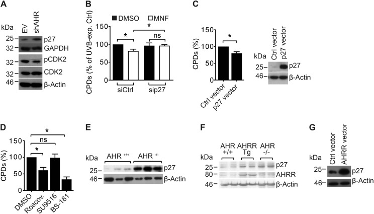
Fig. 2.
AHR inhibits GGR by modulating the protein level of the tumor suppressor p27. a Western blot analysis of p27, pCDK2, and CDK2 in untreated HaCaT-EV and HaCaT-shAHR KC (representative blots). b HaCaT KC were transiently transfected with p27-targeted siRNA and Ctrl. siRNA. After 24 h, the cells were irradiated with 200 J/m2 UVB and treated with 20 µM MNF or 0.1% DMSO. After 4 h, the CPD content was compared by SWB. c HaCaT KC were transiently transfected with Ctrl. vector or a p27 expression plasmid. After 24 h, the cells were exposed to 200 J/m2 UVB and 4 h later the CPD content was determined. d HaCaT KC were irradiated with 200 J/m2 UVB and subsequently treated with 1 µM roscovitine, 500 nM SU9516, 125 nM BS-181 or 0.1% DMSO. After 4 h, the CPD content was analyzed by SWB. e Protein lysates from skin samples of AHR+/+ and AHR−/− SKH-1 mice were analyzed for p27 protein content by SDS-PAGE/western blotting. f Protein lysates from skin samples of AHR+/+, AHRR Tg and AHR−/− B6 mice were analyzed for p27 and AHRR protein content by SDS-PAGE/western blotting. g HaCaT KC were transiently transfected with an overexpression plasmid for rat AHRR or empty vector. After 24 h, the p27 protein level was compared by SDS-PAGE/western blot analysis (representative blot). ns not significant. *p ≤ 0.05

