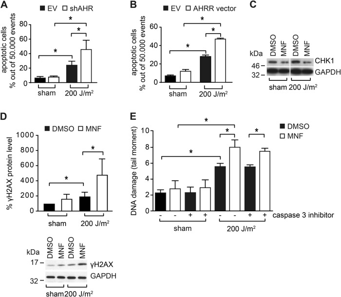
Fig. 4.
AHR inhibition increases UVB-induced apoptosis and is associated with DSB formation. a HaCaT-EV and HaCaT-shAHR KC were irradiated with 0 and 200 J/m2 UVB. After 24 h, the amount of dead cells was analyzed by Annexin V/PI staining. b HaCaT KC were transiently transfected with an expression vector for rat AHRR or empty vector. After 24 h, the KC were exposed to 200 J/m2 UVB and another 24 h later, the amount of dead cells was determined by Annexin V/PI staining. c Western blot analysis of CHK1 in HaCaT KC 24 h after irradiation with 0 and 200 J/m2 UVB (representative blot). d HaCaT KC were irradiated with 0 and 200 J/m2 UVB. After 18 h, γH2AX levels were assessed by SDS-PAGE/western blotting. e HaCaT KC were irradiated with 0 and 200 J/m2 UVB and immediately treated with MNF (20 µM) and DMSO alone or in combination with the caspase inhibitor Ac-DEVD-CHO (20 µM). After 18 h, DSBs were detected by neutral comet assay analyses. *p ≤ 0.05

