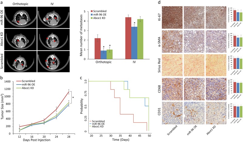
Fig. 4. miR-96 OE and Abce1 KD reduce breast tumor proliferation and lung metastases in vivo.
a Lung CT scans performed on day 28 (orthotopic) and day 21 (IV) post-4T1 injection (left) and quantification of LMets in microCT (right) show significantly fewer metastatic growths (indicated by red arrows) in mice that orthotopically received miR-96 OE or Abce1 KD cells compared to the scrambled control. IV injection of miR-96 OE cells resulted in fewer lung foci compared to Scrambled. b Primary tumor volume analysis showed reduced tumor volume of miR-96 OE and Abce1 KD groups compared to Scrambled c Kaplan-Meier survival analysis demonstrated increased overall survival of mice that received miR-96 OE or Abce1 KD cells compared to the scrambled control (p = 0.06). d Representative images of Ki-67, α-SMA, Sirius Red, CD68, and CD31 staining in resected tumors from Scrambled, miR-96 OE, and Abce1 KD groups as indicated. At least 30 fields were analyzed from each group. n = 3 for each group. Scale bar = 100 μm. Magnification ×20. Quantifications of staining are presented as percent relative to control. miR-96 OE and Abce1 KD tumors demonstrated decreased proliferation (Ki-67) and were associated with significant reduction in activated αSMA + cancer associated fibroblasts in the tumor microenvironment, while collagen deposition (Sirius red staining) was not altered. Data are presented as mean +/− SEM. *p < 0.05

