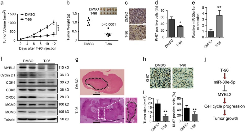
Fig. 7. The anti-tumour effect of T-96 was evaluated in vivo.
a, b Human LN-229 cells were subcutaneously injected into the right flanks of female nude mice. When tumours reached a certain volume, T-96 (30 mg/kg) was injected once every two days, and DMSO injections were administered as the control. Two days after the first injection, tumour volume was measured every 2 days. Two days after the sixth injection, the mice were killed, and tumour weight was measured. c Heamatoxylin and eosin (H&E) staining of the indicated xenograft tumours. d Effect of T-96 on cell proliferation in subcutaneously transplanted tumours derived from LN-229 cells. e qRT-PCR was performed to evaluate the expression of miR-30e-5p in tumour tissues treated with DMSO or T-96. f Western blot assays were performed to evaluate the expression of MYBL2, cyclin D1, CDK4, CDK6, ORC6, MCM2 and MCM3 in tumour tissues treated with DMSO or T-96. Tubulin was used as the control. g Orthotopic implantation was performed with LN-229 cells, and representative images of the heamatoxylin and eosin (H&E) staining are presented; scale bar = 1 mm. h Immunohistochemical staining for Ki67 in tumour tissues. i Effects of T-96 on tumour size (left) and LN-229 cell proliferation (right). j Diagram of the mechanism by which T-96 inhibits cell proliferation and tumour progression in human glioma cells. All data were analysed using unpaired Student’s t-tests and are shown as the means ± SD. *p < 0.05, **p < 0.01, ***p < 0.001

